How X-Rays Changed Dentistry


In centuries past, dentists did not have a way to see what dental issues were happening below the gum line. They could only guess at what was causing a patient tooth pain or discoloration. However, that all changed in 1895 when the x-ray was discovered. With the introduction of this new light wave, dentistry was about to make amazing leaps and bounds in the discovery of dental cavities and how patients were treated. Where dentists once had to simply remove teeth, because of x-ray imaging, they can now see the exact size, shape and location of cavities. Find out how that technology evolved and how it is used on you!
Old Dental Techniques
Dentistry has been around for thousands of years. People in 2000 B.C. had dental needs just like people have dental needs today. Did you know that tooth decay—which you know by the name of cavities—is the most chronic, prevalent disease in the United States? That’s because about 92% of the population has tooth decay by the time they reach adulthood. Many have it in their infancy, and it is the literal decay of the teeth that can’t be reversed.
People thousands of years ago had tooth decay, but did not have the type of modern equipment to deal with it. The first dental school in the world wasn’t even established until 1828, with the first dental college opening in 1840. Both were in the U.S., and almost all effective dental technology was discovered or invented after this time. In fact, if you lived before that time, you would likely go to a barber if you were having tooth pain.
Barbers and surgeons were the ones to see for any type of tooth or mouth issue. However, their ability to fix the dental issues were limited. There was simply no way to really tell what was going on inside a patient’s mouth without cutting open the gums or cutting into the tooth. This is why many people simply had a full tooth painfully pulled when a cavity started to cause them pain or infection. It led to many people losing teeth over time or to having complete tooth loss. It is still this way in many countries. However, dental x-rays completely changed the field of dentistry.
Dentistry and the Dental X-Ray
Many aspects of modern dentistry wouldn’t exist if it wasn’t for the discovery of a specific type of lightwave called the x-ray. This is an electromagnetic wave that cannot be seen by the naked eye. It has a short wavelength, which means it carries a high amount of energy. It’s reported that the x-ray was first discovered in 1895 by a man named Wilhelm Rontgen. He was a German physicist and there were already discoveries that had been made with different types of radiation at this time.
Wilhelm ended up discovering a new type of radiation by chance as he was working in his lab one day. He noticed that fluorescent light would show through certain materials, but not others. This was especially true when it came to the human body, as this new lightwave would show through skin, but not bone or teeth. With a bit more testing, the x-ray is what was discovered.
Serious advances in medicine and dentistry came once x-rays were invented. These rays would pass through the body and on films would produce images of the bones and teeth that absorbed x-ray waves instead of having them pass through. From that time forward, the rays were used to see inside the body and inside the mouth. It completely changed a dentist’s ability to see what was inside a patient’s tooth instead of simply guessing at the problem. X-rays also provided a way to find and treat a cavity instead of simply removing a tooth.

Technology in Dental Offices
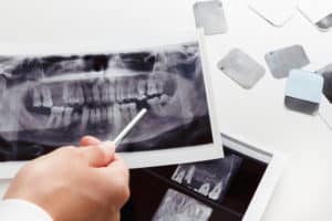
Have you ever had dental x-rays taken at your dental appointment? Odds are that you have. These images are taken at comprehensive exam and dental cleaning appointments. Most patients will have x-rays taken at least once a year, if not at every 6-month check-up. The most common way to take x-rays of your teeth is with bitewings and a moveable x-ray machine. Patients can sit in their dental chairs, and while biting down on a small film of paper, they can have an image taken when the x-ray machine is right by their cheek. In just a few minutes, there are photos of a patient’s teeth.
Imaging doesn’t stop there though. Now, in many dental offices there is a Dental Cone Beam CT machine. This machine allows a patient to stand upright while resting their chin right on the machine. Similar to an actual CT scanner, the machine will rotate around a patient’s body, focusing on the head and dental structures. In just a few seconds, we can get images of your teeth, gums, jaw structures, nerve pathways and soft tissues of the mouth. Without dental x-rays, your experience as a dental patient would be vastly different. However, with x-rays, we have the ability to find cavities when they are very small and treat them. To get your x-rays and comprehensive dental exam, call Dr. Ania’s office today at 303-443-0998!